
There is a good reason eye health practitioners implore that you keep up your scheduled eye exam appointments. It is not to just make sure your eye lens prescription is adequate, it is also essential to make sure you do not go blind. A thorough eye exam can look for and rule out an entire host of macular degeneration marker possibilities easily displayed under a microscopic screening.
Now, retinal calcifications, the formation of tiny spheres made from the same material bones and teeth come from, are another clue that can be added to the search.
Early Triggering Mechanism
Inspecting the retina as you age tells a practitioner your vision health and its possible progression. However, unlike in the past when your eye doctor may have announced that you have indications of this or that and that it was completely normal, many of these minor presentations are now being taken more seriously.
Retinal calcifications were rarely detected in past eye exams. But with new magnifying technology and fluorescent dyes these calcium deposit spheres are now emerging. Rather than dismiss them as minor age-related growths they are now being considered beyond a normal occurrence as a possible early triggering mechanism for age-related macular degeneration (AMD). AMD is currently treated with vitamins for macular degeneration.
In a study reported by the Proceedings of the National Academy of Sciences (PNAS), it was stated that,
“Age-related macular degeneration (AMD) may be caused by deposits of microscopic calcium phosphate spheres in the eye. In the study, [the researchers] note that this is the first time these mineralized calcium phosphate spheres have been implicated in AMD. The possible involvement of these tiny calcium spheres, also known as hydroxyapatite (HAP), could ultimately lead to early detection of the disease.”
Drusen Development Discovery
AMD is often diagnosed through the detection of fat and protein deposits called drusen. These drusen act as barriers of light needed to reach sensitive cells called photoreceptors which assist in creating healthy vision. To make matters worse, photoreceptor cells are recycled through a natural cellular process that creates microscopic waste. It was found that the drusen deposits trap this waste resulting in more deposits in the eye but this time directly on the retina.
Prior to this discovery scientists could not figure out how drusen developed and grew to such a light blocking size but now these tiny calcium spheres, also known as hydroxyapatite (HAP), could be the culprits. In addition, these retinal calcifications are showing possible links to slowly attracting proteins and fats which, over time (years to even decades) eventually form on the surface. This process is now believed to be one of the major causes of age-related macular degeneration and is a discovery considered a breakthrough in combating this sight robbing disease.
According to the PNAS report,
“The researchers made their discovery through post-mortem examination of 30 eyes from donors between 43 and 96 years old. Using fluorescent dyes, they were able to identify the miniature calcium spheres. They also examined tissue samples from AMD patients using X-ray diffraction and fluorescent staining chemicals, which helped reveal the role of HAP in the process.”
Dr. Imre Lengyel senior lecturer and researcher at the School of Medicine Dentistry and Biomedical Sciences at Queen’s University Belfast and lead investigator of the study commented,
“We found these minuscule hollow spheres inside all of the eyes and all the deposits that we examined, from donors with and without AMD,…Eyes with more of these spheres contained more drusen. Our research revealed that early changes in the back of the eye can lead to the build-up of hard mineral deposits, made of calcium and phosphate that may incorporate other types of trace metals, like magnesium. The build-up of these mineral deposits are an indicator of irreversible damage of the retina.”
This study was a joint effort by researchers from a variety of institutions which included: The University of Maryland School of Medicine; University College London; Imperial College London; University of Tübingen, Germany; George Mason University, Fairfax, VA; and The University of Chicago, IL.
This advanced, global collaboration shows how diligently researchers are working to save the sight of so many suffering worldwide, including nearly 10-15 million Americans annually.
PNAS concluded that,
“Dr. Lengyel noted that the spheres appear long before the drusen itself becomes visible during a clinical examination, and that the new techniques can be used to identify drusen buildup long before it becomes visible using current techniques. The findings could potentially advance AMD diagnoses by at least a decade, she added.”
What To Look For
There are some things you can do to help possibly prevent systemic calcifications in your body. These deposits are nothing new as they have often been associated with forming plaque in the arteries which could eventually lead to cardiovascular disease known as atherosclerosis. However, this is the first time such calcifications have been detected on the retina and associated with the development of drusen.
There may be some indications of high calcium levels in your body that may be associated with chronic (constant) symptoms such as:
These symptoms may indicate high calcium levels and if you struggle with these on a daily basis for an extended period of time it is probably best to see your medical or naturopathic doctor. However, in addition, now that researchers have discovered such deposits on the retina it is recommended to have your eyes checked as well.
According to Dr. Curcio, Professor in Ophthalmology at University of Alabama,
“By fully understanding the causes behind the changing environment in which these large, damaging nodules grow we could design new ways to intervene with their growth earlier in the disease process than is currently possible. Identification of these risks associated with disease progression in the eye, especially in the retina, could become a diagnostic tool for monitoring the progression of retinal degeneration. This allows ophthalmologists to counsel their patients more wisely and also allow us to think about slowing or halting the progression of disease, earlier in its course.”
Reduce Potential Calcifications In Your Body
Although calcium deposits can form regardless of a person’s healthy lifestyle, it has been shown that maintaining a high plant-based diet often helps most avoid such developments. It has been found that by consuming a daily supplement along with a diet rich in vitamin K2 most people could reduce a good amount of potential, systemic calcification.
Livestrong reported that,
“Most K2 products come in either 30- or 90-microgram capsules to be taken once daily or in soft-gel form and contain a form of K2 called menaquinone-7, which is readily assimilated and long-lived in the body. The Linus Pauling Institute at Oregon State University suggests “taking a multivitamin-mineral supplement and eating at least 1 cup of dark green leafy vegetables daily.”
It is important to note that you should check with your medical or naturopathic doctor before taking a K2 supplement to make sure it doesn’t contraindicate with your health history or current pharmaceutical protocol.
Stay on top of your sight by maintaining eye exams and paying attention to any changes. In addition, following a plant-based diet and good exercise program could help you avoid developing AMD as well as other health challenges.
 Why Full Body MRI Scans Are Changing Preventative Health
 Sprouts May Help Fight AMD
 5 Foods to Avoid if You have Eczema
 Treating DOMS aka Muscle Fever
 Benefits of Using Skin Toner
 Body Dysmorphia: A Growing Concern
  Achieve Your Fitness Goals Like a Pro with the Best Fitness Benches
 Finding the Best Countertop Water Filter for Safe and Clean Drinking Water!
 Climate Change Skin Defense
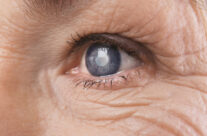 4 Natural Ways to Prevent Cataracts