

As much as researchers continue to probe and scan various organs to find clues of pending disease, sometimes there is just not enough information to see the full spectrum. When it comes to macular degeneration, the eye disease that afflicts upwards of 200 million people globally and continues to rob millions of their sight each year, being able to study the deep effects of this disease has been difficult. The reason is that, until now, only a rodent model could be studied in detail along with two dimensional drawings, scanned MRI’s, and computer generated representations.
Now, researchers have been able to construct a 3-D model breakthrough that advances macular degeneration research. It is such a detailed version that it could enhance investigation and treatment in a more rapid timeframe than it might take otherwise. This could be just the thing researchers across the globe need for easy to follow access that could never be applied before.
Imagine, rather than inject or surgically manipulate a rodents’ retina, this 3-D model offers a near perfect human retina that reacts right down to the nerve and capillary level. This means that procedures and pharmaceuticals could be tested without any need for invasive studies on an actual human. Research this detailed makes all other attempts an antiquated approach as more labs will use this model to enhance the search for an age-related macular degeneration disease (AMD) cure.
Three Dimensional Matrix
Being able to re-create any organ function takes enormous technical and biological skill as the intricacies of everything from minute capillary systems to detailed musculature prove daunting. Researchers at the University of Rochester decided to attempt a functional 3-D model of the human retina. What ensued is a perfect mimic that may be able to predict a lot more than we already know about macular degeneration.
The University of Rochester News Center reported on the makings of this 3-D model.
“Their model combines stem cell-derived retinal tissue and vascular networks from human patients with bioengineered synthetic materials in a three-dimensional “matrix.” Notably, using patient-derived 3D retinal tissue allowed the researchers to investigate the underlying mechanisms involved in advanced neovascular macular degeneration, the wet form of macular degeneration, which is the more debilitating and blinding form of the disease.”
This “three dimensional matrix” opens a whole new world of not only future research but by also answering past questions as well as testing drug efficacy.
Going Deep
One of the biggest speculations amongst macular degeneration researchers is whether the disease is caused by an anomaly of anatomical defects in the retina or by outside systemic issues such as constricted circulation. The 3-D model may be able to answer these long debated questions which looks like the research is currently showing to be anatomical defects. In addition, one of the most immediate effects of using the model is how the choriocapillaris layer has presented.
The choriocapillaris layer is a network of capillaries that brings blood to the outer retina beneath the RPE (retinal pigment epithelium).
According to Ruchira Singh, an associate professor of ophthalmology at the University’s Flaum Eye Institute, the choriocapillaris layer is “a mystery that nobody has ever been able to model in culture,” Creating the 3-D model is, Singh continues, “to get the entire complex that is affected by this disease, so that properties of each individual cell type can be controlled independently,… You can have completely normal choriocapillaris, but if your RPE’s are dysfunctional it will cause the choriocapillaris to dysfunction,”
This preliminary research using the 3-D model is a small example of how scientists can go deeper into the human optical structure than ever before. This immediately surpasses “real-time” image scans or inserted cameras invasively probing and risking infection of human tissue.
Research Underway
Recent results from using the 3-D model have proven to bring newly discovered data on how macular degeneration may form directly under a dysfunctional RPE.
Singh reports the benefits of collaborating with the 3-D model to be:
Dr. Singh collaborated with the lab of Danielle Benoit, professor of biomedical engineering and director of the Materials Science Program located just across the avenue of the University of Rochester. This was a longtime coming as these two women forged a working relationship based on their desire to practice in such an advantageous location. The team, in essence, is a serendipitous collaboration that is the stuff breakthrough science needs more of.
As resources continue to advance, less lab rodents may be needed and more very close to realistic models could become the necessary standard. Rodent research has proved difficult, namely because the anatomy of a human far surpasses that of a rodent. This 3-D breakthrough advances macular degeneration research as well as paves the way for other applications in the future.
Sources:
 Why Full Body MRI Scans Are Changing Preventative Health
 Sprouts May Help Fight AMD
 5 Foods to Avoid if You have Eczema
 Treating DOMS aka Muscle Fever
 Benefits of Using Skin Toner
 Body Dysmorphia: A Growing Concern
  Achieve Your Fitness Goals Like a Pro with the Best Fitness Benches
 Finding the Best Countertop Water Filter for Safe and Clean Drinking Water!
 Climate Change Skin Defense
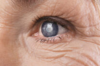 4 Natural Ways to Prevent Cataracts