
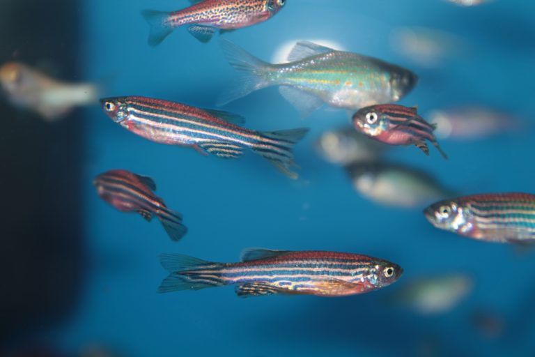
Belonging to the minnow family, one tiny fish with sparkly blue stripes across its translucent body is known as the zebra fish. This fish is very special to medical science as it can easily be researched for various responses to diseases as well as medications. However, recent studies have shown some beneficial results when determining another factor the zebrafish holds, its ability to regenerate retinal neurons.
Researchers have targeted the regenerative ability of the zebrafish which may assist in developing a remedy for the debilitating ocular disease, macular degeneration. Regenerating retinal neurons has not been seen in any living creatures other than this colorful, one-inch bony fish and some of its minnow cousins. Because of its laboratory history, the zebrafish is the most accessible to use for macular degeneration research.
Spontaneous Regenerative Response
The more that humans are able to study the complex systems of creatures from insects right up to large mammals such as whales, wondrous anatomical secrets continue to be revealed. When neurons are lost or damaged by disease or trauma, mammals cannot regenerate them. Once they are compromised they are gone forever. That is, unless you are a zebra fish.
Zebrafish are the perfect specimens for ocular study. Described here in a peer reviewed report prepared by researcher Maria Irbibarne of the Okinawa Institute of Science and Technology Graduate University and published by the scientific journal IntechOpen,
“Zebrafish are small tropical fish that are easy to maintain, and that produce many eggs. They have transparent embryos that develop very rapidly, with a 3–4-month generation time. Zebrafish are also easy to modify genetically. The visual system is highly conserved. It is already functional just 5 days post-fertilization, and it can be assessed by OKR [optokinetic response – ability to follow a moving object]”
In the lab, when these little vertebrates are exposed to optical neuron damage, it has been discovered that a spontaneous regenerative response is triggered. This incredible transformation is the stuff of science fiction as over a short period of time it restores both retinal structure and function. The cells and framework of the retina are very close among vertebrates so the zebrafish fits perfectly into a laboratory aquatic creature to eventual human trial capability.
When it comes to age-related macular degeneration (AMD), if this spontaneous regenerative response in the zebrafish can be harnessed, there may be good news in the near future. It is very possible that an injection of regenerative cells could be delivered directly into the retina to reverse AMD. This would surpass stem cell applications that are just beginning to gain traction but have not yet been perfected for mainstream application. In essence, the zebrafish spontaneous regenerative response skips the step of creating stem cells as the zebrafish does it on its own.
Cellular Activity
Vanderbilt University Medical Center (VUMC) Reporter describes recent study results of longstanding zebrafish AMD research. The study was conducted by Dr. James Patton, Professor of Biological Sciences along with colleagues in the Department of Biological Sciences at Vanderbilt University.
Dr. Patton has been researching the zebrafish retina for several years and has only recently found a significant connection. Patton and his team discovered in the zebrafish a microRNA, known as miR-216a, which regulates the expression of Dot1l, an enzyme involved in regulating gene expression. When the researchers suppressed the miR-216a, Müller glia (MG) cells formed. MG cells are the most common cells found in the human retina and are the cells responsible for the zebrafish retinal regeneration.
Dr. Patton commented to VUMC,
“Müller glia constitute an adult stem cell in the zebrafish retina and our goal is to identify pathways and genes that could be activated to induce similar behavior in the human retina,”
Known as “architectural support,” the Müller glia cells undergo a regression process called ‘dedifferentiation’. This is when they literally and automatically wipe themselves clean of all previous ‘data,’ going from the specialized state they grew into, back to a simpler, more pre-pubescent state. It is essentially a natural stem cell creation that can repair nerve damage like never seen before. When it comes to macular degeneration, this process shows great promise in one day enabling humans to do the same.
The GABA Link
In 2017, Medical News Today reported on Dr. Patton’s early studies of zebrafish retinal regeneration.
The studies involved gamma-aminobutyric acid (GABA), a neurotransmitter that, through chemical messages, regulates communication between brain cells and the nervous system. GABA neurotransmitters are highly prevalent in occurring between thirty and forty times per synapse. However, it was reported that,“high levels of GABA in the retina keep the Müller glia inactive. When retinal GABA levels decrease, the glial cells start to dedifferentiate and then proliferate [increase rapidly], as part of the regenerative process.”
Dr. Patton commented on this process, stating,
“The prevailing belief has been that the regeneration process in fish retinas is triggered by secreted growth factors, but our results indicate that the neurotransmitter GABA might initiate the process instead […] Our theory is that a drop in GABA concentration is the trigger for regeneration. It initiates a cascade of events that includes the activation of the Müller glia and the production of various growth factors that stimulate cell growth and proliferation. If we are correct, then it might be possible to stimulate human retinas to repair themselves by treating them with a GABA inhibitor.”
This previous research shows the painstaking work scientists commit to for a successful result years down the line.
Now that Dr. Patton and his team have been able to identify the GABA link leading to the miR-216a and Müller glia cell manipulation, research may advance more rapidly. This is at least the hope as human trials often present a host of unexpected and unexplained responses. However, given the fact that the zebrafish ocular structure is a good comparison to the human eye, the hope is that these discoveries will offer a seamless crossover for macular degeneration treatment.
 Why Full Body MRI Scans Are Changing Preventative Health
 Sprouts May Help Fight AMD
 5 Foods to Avoid if You have Eczema
 Treating DOMS aka Muscle Fever
 Benefits of Using Skin Toner
 Body Dysmorphia: A Growing Concern
  Achieve Your Fitness Goals Like a Pro with the Best Fitness Benches
 Finding the Best Countertop Water Filter for Safe and Clean Drinking Water!
 Climate Change Skin Defense
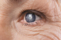 4 Natural Ways to Prevent Cataracts