
Age Related Macular Degeneration is the leading cause of blindness and central vision loss in adults over the age of 65. It is estimated that 10-15 million Americans suffer from AMD. As we age, the macula, the integral part of the retina responsible for vision, begins to deteriorate.
Yellowish deposits called drusen accumulate on the macula damaging the light sensitive tissues, leading to blurry vision and often impaired day to day living. This is known as dry AMD.
The wet form of macular degeneration is the result of leaking blood vessels on the macula. The wet form is much more concerning because approximately 80-90 percent of blindness associated with macular degeneration is from the wet form.
If the wet form is left untreated for too long, it can result in irreversible damage and vision loss.
The challenge for eye care specialists has been determining the likelihood of when one may progress from the dry form to the wet form. Seeing as the current treatment options for wet AMD are both costly and invasive, physicians may not start a patient on a treatment if they are in the intermediate stages of the disease.
This leaves the timing of their next checkup crucial for catching the progression and leaves doctors and patients hoping that the next visit will be soon enough to prevent significant damage.
 In a study published in the November issue of Investigative Ophthalmology and Visual Science, researchers lead by Daniel Rubin of Stanford, have discovered a way to predict the probability of a patient’s dry AMD progressing to the more serious wet AMD.
The method used to predict whether one will progress from the intermediate stage of AMD to the wet stage is based on a formula which crunches data from routine retinal scans performed by optometrist and ophthalmologists.
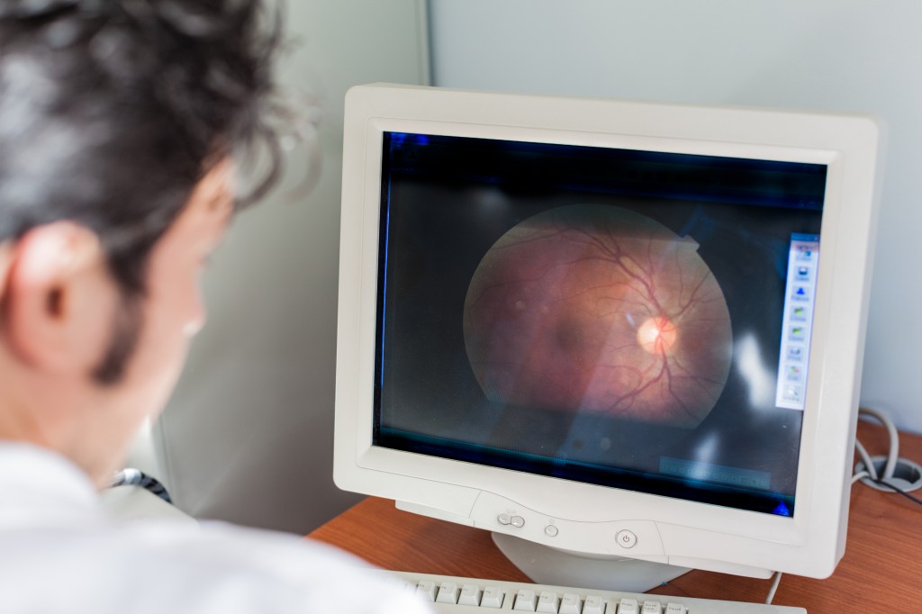
“Right now, a patient who goes into the ophthalmologist’s office typically gets an SD-OCT scan anyway,” said the study’s senior author, Daniel Rubin, MD, assistant professor of radiology and of biomedical informatics. “Our technique involves no new procedures in the doctor’s office — patients get the same care they’ve been getting anyway. We’ve simply added on a computerized image-processing step that analyzes not only that scan but any previous ones available from that same patient’s earlier visits.”
The scan, which is very similar to an ultra sound, focuses a laser beam at the macula. The reflecting light from the macula is converted into data points, which in turn are converted into a 3D, high resolution image of the eye.
Upon analysis of the scans physicians are able to determine a risk score which can predict the likelihood and more specifically time frame in which one’s disease may progress.
This is a vast improvement since previously doctors didn’t have a way to generate a risk score. The risk score allows doctors to predict how long it will take for someone to progress to the wet stage. For example, they can predict 1 year, 2 years, 3 years etc.
This allows for a concrete method of scheduling follow up office visits particularly for those with a probability to progress within 1 year.
The study followed 244 patients seen in Stanford Health Care over a five year period and the model accurately predicted each case of progression within a year, yet 40 percent of the time the model predicted progression within a year, it did not happen.
“No test gets it right 100 percent of the time,” Rubin said. “You can tweak the model to trade off the risk of telling someone they will progress when they actually won’t against the risk of telling them they won’t progress when they actually will. With AMD you really don’t want any false negatives, so you tune the model accordingly. The downside is that some patients will wind up being told to come in sooner than, in fact, they probably need to. But that’s nothing compared with the downside of a patient at high risk for progression not coming in soon enough.”

Exposing the Hidden Danger to Your Eyes Do you know the #1 WORST food for y...

Getting enough sleep is challenging for many people. When you are pressured...

It's long been common knowledge that getting enough potassium every day is...

Best resource for tips to buy best macular degeneration eye vitamins and su...

If you are like many of us that are making efforts to eat a healthier diet...

Located in back of the abdomen and about the size of a fist, each of your k...

You may have heard of all the hype about taking vitamin D but might not be...

What if some of the worst degenerative diseases known to man didn't have to...
Your fingernails are front and center yet few people really inspect their o...
At one time or another you must have found yourself biting your fingernails...
 5 Foods to Avoid if You have Eczema
 Treating DOMS aka Muscle Fever
 Benefits of Using Skin Toner
 Body Dysmorphia: A Growing Concern
  Achieve Your Fitness Goals Like a Pro with the Best Fitness Benches
 Finding the Best Countertop Water Filter for Safe and Clean Drinking Water!
 Climate Change Skin Defense
 4 Natural Ways to Prevent Cataracts
 Move More, Live Longer
 Vegan Ice Cream Review and Recipe